|
The Optivision CornealMap EH-300 is the result of 40 years of
research and development. It is the descendant of the first fully automated Corneal Topography system. The Optivision CornealMap
EH-300 introduced the following innovations to the field of Corneal Topography:
1) Auto-positioning/ auto-centration/
auto-focusing
2) Tangential/ Sagittal/ Gaussian/ refractive displays
3) Determined the X,Y,Z coordinates for
each point on the cornea
4) 3-D display
5) Elevation maps (the first Z maps was shown at the AAO meeting in
Las Vegas, 1987)
6) Simulated fluorescein contact lens fit
7) ASCII file (output data)
8) Side by
side patient doctor (operator)
9) Keratoconus detection
10) Wavefront measurements of the cornea with Zernike
polynomial
11) Fourier analysis with display of the harmonics
12) Summary plot showing the tangential, refractive,
elevation and wavefront plots
13) Difference plots for dioptric, refractive and elevation
14) Innermost area measured
.46mm, outer measurement 10mm plus
15) Microsoft Windows XP
16) Integrated e-mail system for transfer of data files
What is the Science behind todays topography systems?
There are numerous corneal topography systems available
today. One system, the CornealMap EH-300 Corneal Topography System, handles and displays data in a unique way, providing three
levels of information. The CornealMap system provides corneal curvature in three ways, tangential (shape), sagittal (power)
and Gaussian. Moreover, using the Z coordinates, the CornealMap can display the Wavefront of the cornea using the Zernike
polynomial or the Fourier analysis to decompose the wavefront in separate harmonics.
The Optivision CornealMap measures
the X,Y,Z coordinates of each points on the 23 rings and 360 meridian and up to 8280 locator points to map the cornea
The relationship between the cone, rings, optical axis, and camera of the CornealMap system is known by design and fixed
in manufacturing. The CornealMap system locates the apex and the imaginary plane that touches the cornea at the apex. Scanning
the digitized image will provide the x and y coordinates for any location. The distance between each point can be measured,
and, since distance is due to curvature in the cornea, the Z coordinate for each point can be calculated. The 23 rings over
360 meridians give 8280 locator points on the corneal surface, providing greater coverage and sensitivity to the shape of
the corneal surface.
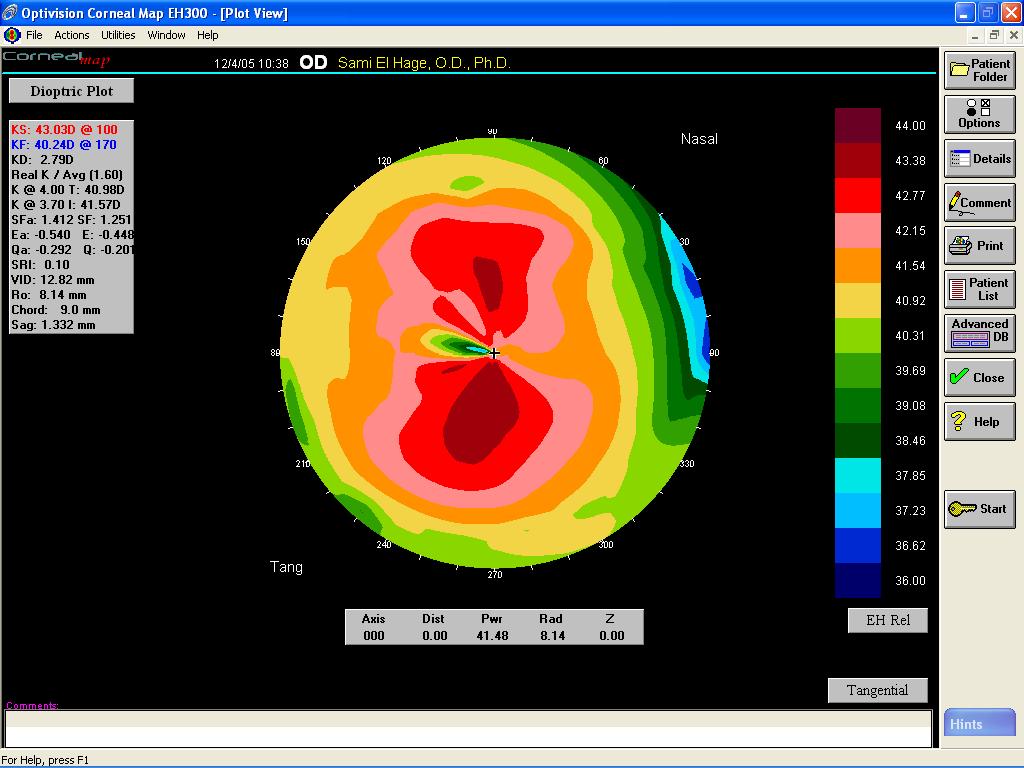
By using the X,Y, and Z data, a 3D image is generated, demonstrating the changes in curvature across
the surface of the cornea. This three-dimensional plot is an excellent tool to educate patients. It makes it easier for them
to understand the image without having to know what the colors mean. The most popular display is the dioptric plot, a view
of corneal curvature in terms of power, orientation and astigmatism.
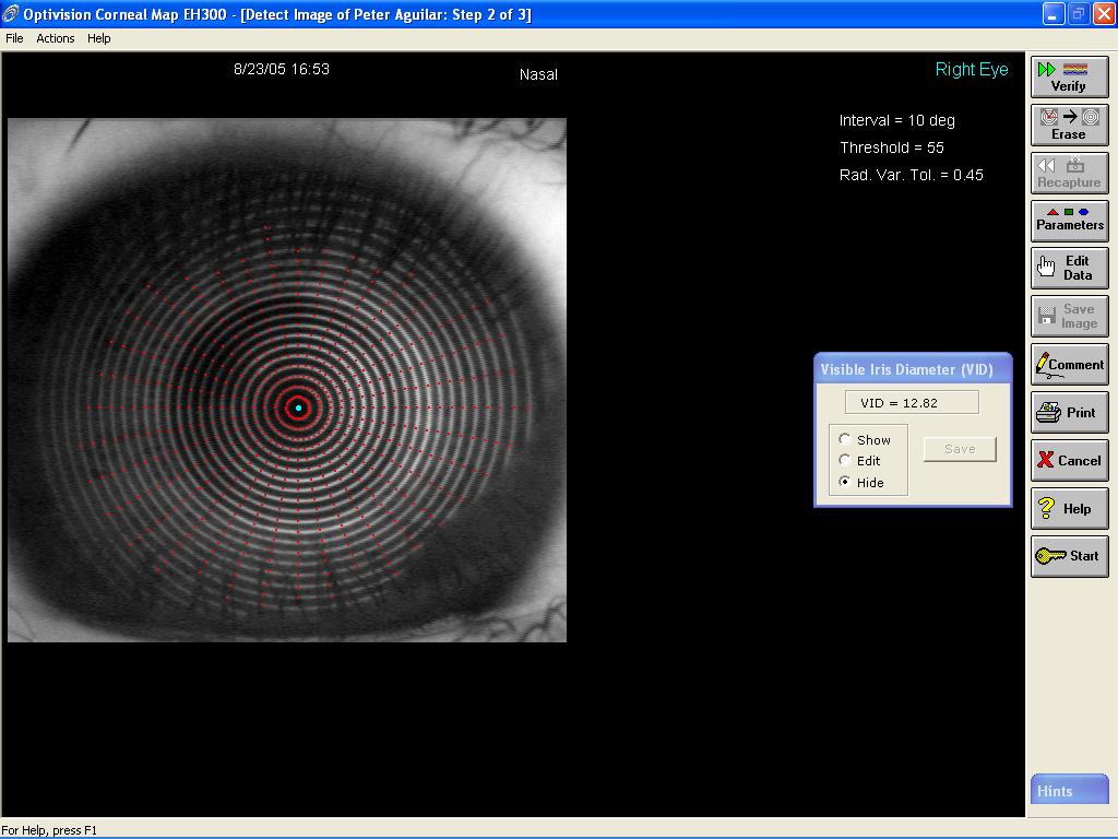
Automatic Centering, Focusing, and Capturing
The CornealMap EH-300 system, now in its fifth generation, was the first system to automate centering, focusing, and capturing,
a most helpful feature. The CornealMap can now capture up to five consecutive pictures. When different operators are introduced
to the equation of patient image capture, some assistants bias the focus to their preference. This automated function assists
in eliminating operator bias, which is imperative for accurately capturing the data. The feature speeds up exams and ensures
compatibility among operators. For difficult patients, however, the CornealMap system incorporates a manual image-capture
override to allow the operator to capture an image manually.
Once the image or images has been captured and locked into
the digital memory, the operator can now examine the quality of the mires using the analysis to ensure that data collected
are good. The operator can excludes at will the out of focus image and keep the rest for measurement. An average plot along
with the other plots is displayed.
Sensitivity is key, especially in PRK
The inner ring of the CornealMap
EH-300 system is .46mm diameter, and the outer ring is 10 mm, allowing for a great sensitivity of the central corneal surface.
This feature is presently important but even more useful now with excimer refractive surgery. Following refractive surgery,
some of the visually significant corneal irregularities are very subtle and easily missed. Central islands are residual, steepening
in the center of the refractive surgery ablation. Competitive systems may miss these islands and only show a smooth central
surface. Diagnosis of such irregularities will be paramount in postoperative care of Lasik/Lasek/PRK surgery patients.
Analyze images in 10- and 1- degree steps
The Optivision CornealMap EH-300 system also has the capability of analyzing
an image in 10- and 1-degree steps. The 10-degree detection variation offers sufficient analysis of normal corneal surfaces.
When the subtle details of incision sites, transplant margins, or contact lens warpage are needed, the 1- degree detection
will provide the most data and coverage. When small epithelial irregularities cause overlying tear film breakup that can affect
results, the 10-degree mode eliminates erroneous data from the map.
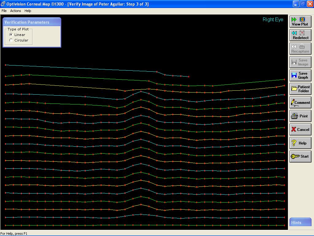
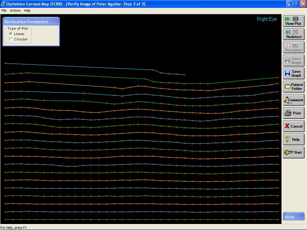
Data Verification
One of the many special
features of the CornealMap EH-300 system is the Verify window. The Verify window provides a guide to accurate analysis of
the data. The function eliminates technician subjectivity, because it verifies the accuracy of the image captured and determines
whether the data are dependable and true. The competitive systems require close scrutiny of the data points on the image to
determine whether the data are useful, encouraging operator subjectivity. Without the CornealMap EH-300 Verify window, other
systems allow analysis of data that should have been discarded and recaptured.
|
|
Why is networking/co-management important?
An important
consideration for eye doctors is the ability to participate in the new explosion of Surgical Keratorefractive referral and
co-management organizations. Ophthalmologists and optometrists alike will be interested in a corneal topography system that
has the capability of sending patient data, images, and graphs to another offsite doctor or center: The CornealMap EH-300
system has software capable of achieving this.
Send data anywhere
The receiver of the patient files will
be able to view or manipulate and alter the data using CornealMap system or CornealMap EH-300 Executive Software. A single
CornealMap EH-300 or multiple CornealMap EH-300 systems are connected to a local area network (LAN) and Internet E-mail service
provider. Remote locations on the LAN and those with a sending/receiving E-mail address can store and retrieve patient files.
The method for sending the data to remote sites uses any Windows-compatible E-mail client. The Windows software facilitates
system operation and patient file management. This will expedite as well as improve patient co-management among doctors at
different sites. It will be helpful with expediting second opinions and advising doctors responses.
|
Advanced design
The design of the CornealMap EH-300
system enables the operator to position the patient for the optimum use of office space. The components can be arranged side
by side, allowing the assistant to directly visualize head position and assist, or if necessary, face to face 90 degrees,
providing flexibility and patient comfort. The feature is convenient for operator and patient. The system is even designed
to comfortably accommodate patients using wheelchairs.
Is there technology that can help in difficult-to-evaluate
cases?
The CornealMap EH-300 technology provides the flexibility to evaluate difficult corneal cases that fall outside
of the standard range. Because the Optivision CornealMap Topography system has good corneal coverage and a wide range of adjustable
parameters, almost any irregular corneal surface can be imaged and analyzed.
|